Although invisible to the naked eye, measuring just 125 billionths of a metre across, the pandemic virus is now an iconic image.
Scan any newspaper or watch any broadcast and you can catch a glimpse of what looks like a vintage mine, the spiked ball of a medieval weapon, or a greyish sphere, sometimes armed with blood-red spikes, other times with yellow or blue protrusions.
The Science Museum Group Collection is rich with objects inspired by attempts to show what the molecules of life would look like if we could shrink to the atomic level.
Famous examples include the model of DNA’s double helix, created by Jim Watson and Francis Crick, and Sir John Kendrew’s ‘sausage model’ of the muscle protein myoglobin (though some, unkindly, refer to it as the ‘dog poo’ model).
But COVID-19 is exceptional.
As Geoff Belknap, Head Curator at the National Science and Media Museum, and former Curator of Art Katy Barrett commented on COVID-19 in a recent special issue of the journal Interface Focus: “No previous virus or disease has gained such visual currency for the image of its source, an image rooted in processes of scientific imaging and illustration.”
I discussed how to visualise the pandemic virus with artist Angela Palmer, along with the scientist-artist David Goodsell.
Angela Palmer
While studying anatomy at the Ruskin School of Drawing and Fine Art in the University of Oxford, Angela Palmer developed a way to create ghostly images of the interior of brain, such as of the TV personality Carol Vorderman and novelist Robert Harris – and of the body too – by engraving details from medical scans on to glass, layer by layer, to create a three dimensional representation.
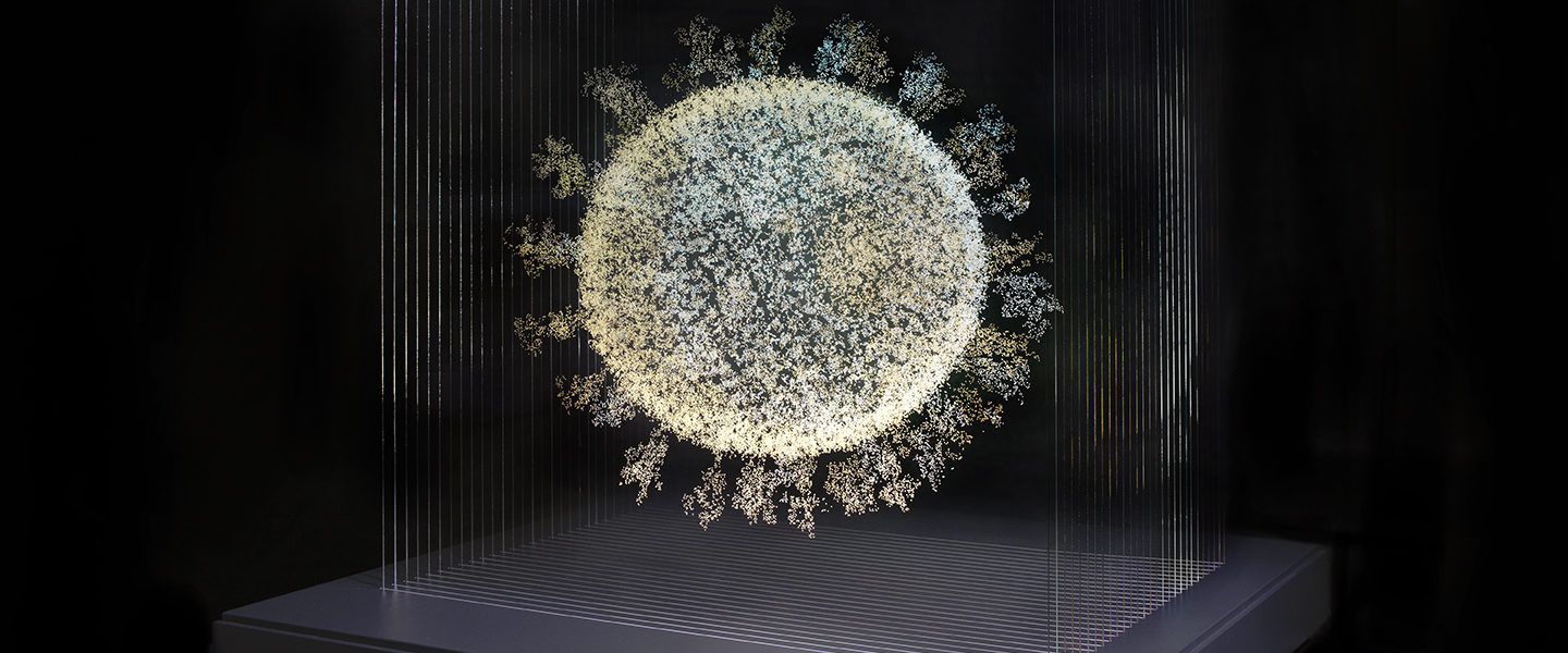
In 2020 she used the same approach with COVID-19, which has been imaged down to the atomic level by various methods, such as cryo electron microscopy, to obtain cross sections of the virus which she then engraved by hand onto glass sheets.

Now the Science Museum Group has acquired 2020: The Sphere that Changed the World. I talked to Angela Palmer, and her edited responses are in italics.
I wanted to re-create the virus particle, as a three-dimensional ‘drawing’ floating in glass, using the same technique I adapted all those years ago, inspired by Dorothy Hodgkin’s model of the penicillin molecule (which can be found in the Science Museum Group Collection).
How did you hope to depict COVID-19?
I am hard wired to reflect on what is happening in the moment. It was all about the hidden enemy that everyone is afraid of, this particle sphere.
My plan was to engrave the details of the virus onto 28 glass sheets, lit from below, similar to the one I created in The Smithsonian Air and Space Museum of the Kepler telescope’s findings (the observatory hunts for planets around other stars, notably those that are potentially habitable).
What images of the COVID-19 virus did you use?
Tracking down cross sections of the virus has been a fascinating quest – the Oxford neuroscientist Irene Tracey (our paths crossed in Oxford) put me in touch with Sarah Gilbert (just a tad busy with the Astra-Zeneca vaccine!) and Elizabeth Garman, professor of molecular biophysics. The latter sent me in the direction of Professor Dmitry Korkin at the Worcester Polytechnic Institute in Massachusetts, who was using molecular modelling of the virus to reconstruct the virus, having previously worked on the Sars virus.
Happily Professor Korkin agreed to collaborate on my project. He had already mapped the protein spike, which is not only very beautiful but as you will know is the key target in vaccine efforts. It therefore struck me that I should create two installations – one of the protein spike to sit alongside the one of the entire virus particle.
How did it feel when you first saw it?
It was an extraordinary moment when it all came together as a sphere. Here is something that is a menace, that we have reviled and been afraid of, that is strangely beautiful, a natural form. It is beautiful, complex and paradoxical. It is the sphere that changed the world.
What do you think visitors will make of your COVID work?
People will see it as trapped, while it in turn trapped humanity.
The sheets, assembled into an ethereal sculpture, may prompt reflection and contemplation of loss – a loss that will of course be unique in form to each observer.
Laid bare, the particle sphere may also offer empowerment and agency, seen suspended and imprisoned in a glass chamber, suddenly solitary, isolated and vulnerable.

David Goodsell
The pandemic virus inspired an unusual effort to blend art and science by computational biologist and science illustrator David Goodsell of the Molecular Graphics Laboratory in The Scripps Research Institute, La Jolla, California.
The effort to visualise the invisible world of the cell goes back to the birth of molecular biology in the middle of the last century, when the molecules of life were revealed by how they scattered X rays.
Today, proponents of what is called structural biology can also use a form of electron microscopy, called cryo-em, to inspect living things, including the SARS-CoV-2 virus responsible for the pandemic, down to the atomic level.
Scientists today explore these hidden worlds on a computer, showing the molecules of life, often decorated in garish neon colours to signify different proteins, the building blocks of cells and viruses, or to distinguish different atoms.
David Goodsell uses computers too, notably to design drug molecules to target proteins in the body and to reveal how proteins densely crowd into the innards of cells, based on knowledge of the structures of proteins in an online database used by scientists worldwide: the Protein Data Bank, a global repository of genetic and structural data on the tens of thousands of the proteins.
However, the way he illustrates these worlds is informed by the older medium of painting. He uses a pastel palate of colour (ultimately, the ‘colour’ of an atom is an artistic choice), so the spikes are now pink rather than blood red.
He prefers gentle colours and curved forms over the stark primary hues and jagged spikes of most coronavirus images. And he likes to use watercolours because ‘computational modelling was not up to creating the images I wanted. The reason I do my paintings is I have a high school student or armchair scientist in mind who wants to learn more.’
In this way, he could render this agent of destruction into a beautiful image, which he released on Twitter. Among the comments he got was ‘how can something so pretty be so dangerous?’
‘A lot of people were using them with their kids as a way to talk about viruses, to show that they have a structure, are real objects and ones we can fight,’ he said. ‘They put a face on the virus. We did a colouring book version so people could play with it themselves.’
That does not mean to say he takes artistic licence: he shows only the known proteins in the virus and how they might be organized.
Viruses are the simplest things that could be considered living (at least when they are exploiting the metabolism of a cell – outside of a cell they are elaborate packages of chemicals).

Now Goodsell has extended his art to an extraordinary project to create a ‘virtual cell’, led by systems biologist Markus Covert at Stanford University.
While a graduate student, Covert was captivated by the first comparative genomics study, when the team led by gene sequencing pioneer Craig Venter juxtaposed the genetic code of the tiny bacterium Mycoplasma pneumoniae and Haemophilus influenzae to begin to work out the core set of genes required for life.
Covert can still remember the excitement of reading an article that cited one of Venter’s team, Clyde Hutchison, who talked about how a working computer model of a cell would be the ultimate test of biological understanding.
That quotation would stay with Covert, whose team used Venter’s sequencing data of Mycoplasma along with data gleaned from more than 900 scientific papers, to create the first model of a bacterium that took into account all known gene functions of its 525 genes.
To reveal the structure of his bacterium, enter David Goodsell. As he remarked, Covert’s data ‘showed us what the pieces are, and where they need to go.’ Using these data, Goodsell’s postdoc Martina Maritan has sought the molecular structures of each individual protein and assembled them into the bacterium over three years with the help of software developed by her colleague Ludo Autin. The beautiful result shows different levels of detail within the simple bacterium, helping scientists to visualise a cell in three dimensions.
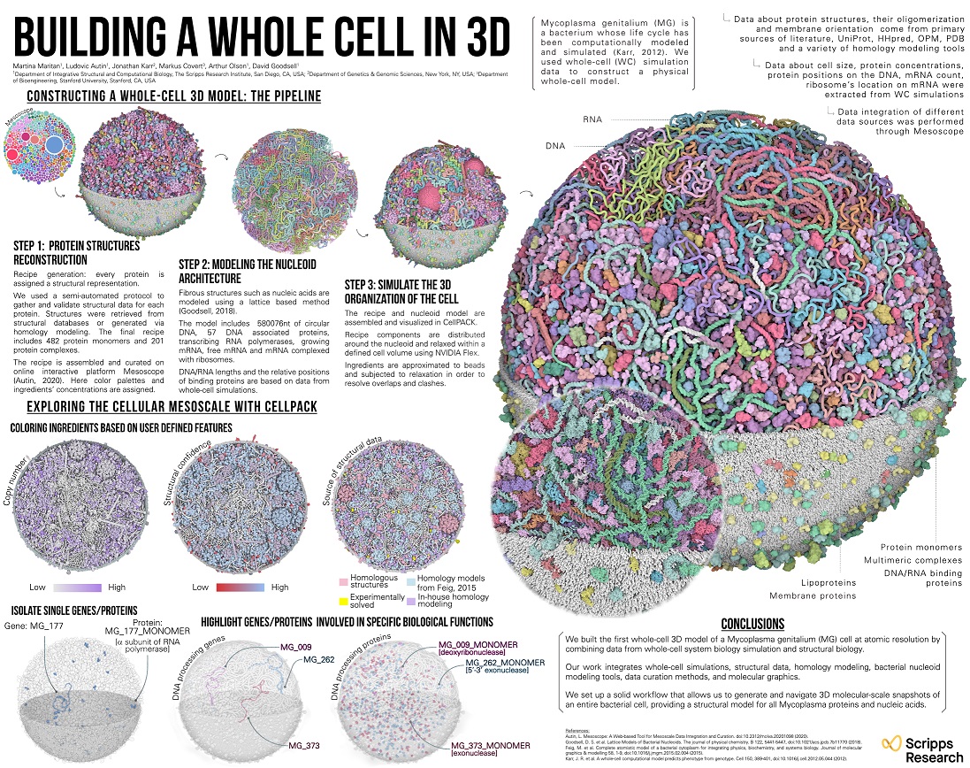
The director of the laboratory, Art Olson, also experiments with virtual and augmented reality, along with 3D printing to help visualise the invisible worlds of molecular biology. Among his creations is a model of a virus consisting of protein pieces and magnets, which self assembles, just like the real thing. ‘You take the 12 pentameric pieces, put in bottle and shake and the whole thing will self-assemble’, said Goodsell. ‘It’s amazing.’
HOW CAN I FIND OUT MORE?
The latest picture of how far the pandemic has spread can be seen on the Johns Hopkins Coronavirus Resource Center or Robert Koch-Institute.
You can check the number of UK COVID-19 lab-confirmed cases and deaths along with figures from the Office of National Statistics.
There is more information in my earlier blog posts (including some in German by focusTerra, ETH Zürich, with additional information on Switzerland), from the UK Research and Innovation, UKRI, the EU, US Centers for Disease Control, WHO, on this COVID-19 portal and Our World in Data.