Where did viruses come from?
Viruses are tiny, consisting of a scrap of genetic code wrapped in fat and protein. When it comes to their ultimate origin, there are many theories.
To explore the origins of viruses, the most abundant biological entities of all, I consulted Jean-Michel Claverie, Emeritus Professor of Genomics and Bioinformatics at the School of Medicine, Aix-Marseille University.
Claverie is a well-known virus hunter who even discovered giant viruses that can ‘catch colds’.
Viruses have been around as long as cellular life itself, which is around four billion years, because they depend on infecting cells to multiply, Claverie explained.
‘The virus-first theories are nonsense’, he said. ‘A straightforward scenario for the emergence of viruses is their evolution from cells, although maybe not the same kind of cell that has come to dominate today’s world. There is also no need to impose a unique or single origin of viruses.’
Viruses can carry their genetic information in the form of the long chain-like chemical RNA (as in the case of the coronavirus), or DNA, and he said we don’t know if RNA viruses are ancestors of DNA viruses or if they appeared independently.
Some believe the first life relied on RNA, though others think RNA has always coexisted with DNA in living things (as is the case in the cells in our own bodies, where RNA carries out DNA’s instructions to build proteins).
Claverie, however, believes viruses ‘are likely very old’ and first originated from ancient RNA cells, though they have emerged many times through ‘reductive evolution’.
This is where the virus evolves to replace as many key steps it needs to reproduce by increasingly relying on the chemical machinery of the cells that it invades.
Reductive evolution has also been seen in parasitic bacteria, he explained, where these microbes have tiny genetic codes and come to rely on host cells for much of their metabolism, save the ability to divide in two and make their own proteins.
However, viruses are even simpler than these bacteria, he said: neither do they make their own proteins – these are manufactured by the cells they infect – and nor do they multiply by cell division.
Instead, they spread their genes by wrapping them in an overcoat of protein and fat, ‘like seeds’. In common with an idea set out in 1957 by the pioneering microbiologist André Lwoff, Claverie believes the way they propagate is a key defining feature of viruses.
Where did the COVID-19 virus come from?
When it comes to the immediate origins of the SARS-CoV-2 virus, analysis of its RNA code links it to viruses that infect bats and the pangolin, a scaly anteater.
Genetic analysis reveals that SARS-CoV-2 is most closely related to a group of viruses found in horseshoe bats (Rhinolophus).
However, unsubstantiated theories — promoted by US President Donald Trump — that it escaped from a laboratory in China still persist.
How did we first discover and understand the nature of viruses?
For a brief history of virology, I talked to my Science Museum colleague, Katie Dabin, curator. Her edited comments are shown below in italic.
When the English doctor Edward Jenner developed the first vaccine to protect against the smallpox virus in 1796, no-one – Jenner included – knew that viruses existed and were the cause of many illnesses. Diseases were explained as acts of God, imbalances of the humours, or caused by miasma’s – ‘bad air’.

It wasn’t until the late 1800s that scientists began to show that something other than and smaller than bacteria were causing plant and human diseases – though these early experiments couldn’t reveal exactly what was at work. Unlike bacteria and other microorganisms, viruses were too small to be seen under light microscopes – the only bit of kit researchers had at that time to detect phenomena too small to be seen by the eye.

The new coronavirus measures about 125 nanometres (125 billionths of a metre) in diameter, compared to about 10 micrometres (one millionth of a metre) for the cells that it invades, and 400-600 nanometres for the wavelength of visible light.
The virus’s genetic code carries the instructions to make just 29 proteins.
How did we detect the first virus?
Tobacco Mosaic Virus (TMV), which infects plants, was the first virus to ever be identified, and has remained at the forefront of viral research ever since. Evidence of the virus emerged in the late 19th century, when a mysterious infectious disease was damaging tobacco crops.
If you’ve seen any of our extraordinary smoking objects you can imagine the risk of tobacco plantations being ravaged by the virus, spelled social and economic disaster. Many researchers began to investigate the invisible agent causing this disease.

In 1892, Russian researcher Dmitri Ivanovsky gave the first concrete evidence that a new agent which was much smaller than a bacterium was infecting the plants using a Chamberland filter, this was cutting-edge technology for that time.
The porcelain filter, invented by French microbiologist Charles Chamberland (1851–1931), had pores so small that only something smaller than bacteria could pass through it. Ivanovsky showed that the plant sap remained infectious, even after it had been passed through the filter, and concluded it must contain a new, never-before detected agent of infection.

Dutch researcher Martinus Beijerinck built on Ivanovsky’s observations and showed that the ‘filtered’ disease agent relied on growing leaves to multiply as it couldn’t reproduce without them.
We know now that viruses are only able to multiply inside the cells of other living things. Beijerinck coined the word “virus” from the Latin for a liquid poison, to differentiate these new filterable pathogens from bacteria.
Ultimately, these early researchers weren’t able to stop the tobacco mosaic virus ruining many plantations but their pioneering work opened the door to viral research that has revealed a better understanding of viruses, their role in human disease, and our ability to tackle them using vaccines amongst other therapies.
Ok, so that’s a plant virus, what about detecting human viruses?
Not long after the discovery of the tobacco mosaic virus, the first ‘filtered’ virus found to cause human disease – Yellow Fever – was discovered in 1901.
At the end of the 19th century, for every soldier who died in battle when the United States invaded Cuba during its war with Spain, 13 died of Yellow Fever.
The disease derived its name from symptoms including the yellowing of eyes and skin, the result of jaundice due to liver damage.
Many researchers concluded that it was transmitted by mosquitoes, but it was American scientist James Carroll who demonstrated filtered blood serum from one patient would induce disease when injected into a second. Yellow Fever became the first human infectious disease to be attributed to a virus.
When did we actually begin to see viruses?
Scientists only began to see what these invisible agents looked like after the invention of the electron microscope in 1931, by Ernst Ruska and Max Knoll. Using electrons rather than light, these microscopes began to transform what could be seen and understood about viruses.
After coating their virus sample in gold nanoparticles (to stop the electrons “burning the sample to ashes”) in 1938, Ruska’s brother Helmut, a medical doctor, saw what a virus looked like for the first time by imaging – you guessed it – the tobacco mosaic virus. Helmut was able to roughly determine its size and showed it to be rod-shaped.
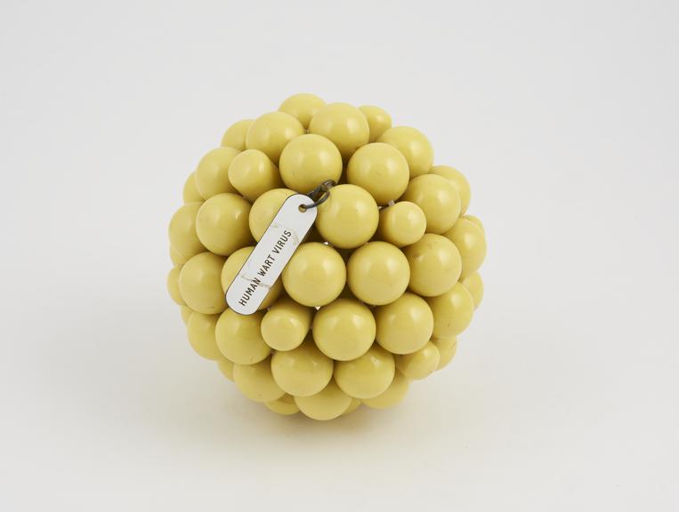
Although limited by the standard of today’s tech, the images the Ruska’s EM revealed that viruses ranged in shape and size, in contrast to TMV, as for example, a mouse pox-virus appeared much rounder.

Detail view of eyepiece of microscope.
Who was the first to study a coronavirus, like SARS -CoV-2?
I have written about June Almeida, the pioneering Scottish electron-microscopist who identified the first coronavirus, and observed its crown-like spike appearance, after which this family of viruses is named.
She published the first photographs of a coronavirus in the Journal of General Virology in 1967. The Science Museum Group collection contains her research papers and teaching materials – including three simple virus models and a collection of glass slides with EM images of viruses.
Does EM tell us everything about the make-up of viruses?
In simple terms, no. To find out more about the building blocks that make up a virus’s structure, researchers pioneered a different technique called x-ray crystallography, to reveal the molecular detail of viruses.
Essentially it involves firing x-rays at crystallised substances and plotting their structure from the patterns those rays make by diffracting or bouncing off the molecules that compose it.
The first step in showing that viruses could be imaged using x-ray crystallography techniques, was to show that they could be crystallised in the first place.
When Wendell Meredith Stanley purified and crystallised the tobacco mosaic virus in 1935, proteins had only been able to be crystallised for a short time. Therefore it was astonishing at the time to discover that a replicating organism could be crystallized.
Researchers began to use x-ray crystallography to try to determine the 3D structure of TMV, but it was the incredible talent of the English chemist Rosalind Franklin in the 1950s that used the technique to reveal the units and helical structure of TMV in unprecedented detail.
More on the 29 proteins of the coronavirus and how they pirate human cells can be found in an earlier blog. I have also discussed the molecular biology of the virus, among other things, with Venki Ramakrishnan, Nobel prizewinner.
Didn’t Rosalind Franklin have something to do with DNA?
Yes! Rosalind Franklin is best known for her x-ray diffraction studies of DNA that provided the vital clues needed by James Watson and Francis Crick to show that the structure of DNA was a double-stranded helix.
However, after her work on DNA, Franklin went on to study the three-dimensional structure of viruses at Birkbeck College in London. This is where she spent the last five years of her life, conducting pioneering work into the structure of viruses.

Why did Franklin study TMV?
TMV was a convenient virus to work with for x-ray crystallography studies. It was readily available and formed crystals easily. Its rod structure suggested it was relatively simple in structure for a virus.
In the 1950s, when computers were novelties, solving the structures of even simple molecules was an immense challenge – and viruses were much more complex assemblies of proteins and nucleic acids.
For Franklin, studying TMV’s structure addressed fundamental questions about the mechanics of living processes – particularly the role of protein and nucleic acid on the activity of living cells.
What did she find out?
In the second observation of a helical structure in her career, Franklin’s x-ray diffraction images revealed the rod-shaped tobacco mosaic virus to be a hollow tube made of proteins. Inside which, there are spirals of a single strand of RNA.
In fact, Franklin built an enormous model of the helical structure of TMV, rivaling that of DNA for the 1958 World’s Fair in Brussels, of which the Science Museum holds an exact replica.
The extraordinary model, representing a segment of the TMV helix, was around 2 meters tall. On 16 April 1958, just as the Brussels exhibition opened to international acclaim, Franklin died of cancer.

What do we know about TMV today?
The protein coat of the virus is composed of more than 2000 copies of a small protein, which stack like bricks in a cylindrical chimney. The RNA strand encodes four proteins, which together, orchestrate the life cycle of the virus.
Once inside the plant cell, the protein coat falls away and the nucleic acid portion directs the plant cell to produce more virus nucleic acid and virus protein. This disrupts the normal activity of the cell.
What is surprising is that unlike other viruses, the tobacco mosaic virus is very stable. So stable that it can survive for years in cigars and cigarettes made from infected leaves.
That contrasts starkly with the SARS-CoV-2 virus, which is round and wrapped in a fatty envelope that makes them especially susceptible to soap when you wash your hands.
Is TMV able to help us in the fight against COVID-19?
Some researchers are exploring whether TMV and tobacco plants can help us meet demands for producing diagnostic reagents and antibodies on a global scale of production in order to tackle the COVID-19 pandemic.
Plants may offer one solution to manufacture these critical ingredients in a timeframe of weeks, compared with months or even years for cell-based systems.
Tobacco plants grow rapidly and can be engineered to produce the relevant antibodies for tests and vaccines. This works by combining TMV with a cloned portion of the genetic sequence encoding a selected coronavirus antigen.
Vaccines or test agents could be harvested and purified from the plants. The idea is undergoing preclinical testing.
It will be interesting to see if a simple plant virus (like TMV) continues to make history within virus research, and in the context of a global pandemic.
what can electron microscopy show us today?
One of my earlier blogs described how electron microscopy has been refined to take images of proteins, using a technique known as cryo-EM. Now for the first time, the technique has been refined further so it is able to discern individual atoms in a protein.
The advance was described by Holger Stark and colleagues at the Max Planck Institute for Biophysical Chemistry in Göttingen, Germany, and a team led by Sjors Scheres and Radu Aricescu at the Medical Research Council Laboratory of Molecular Biology (MRC-LMB) in Cambridge.
Ultimately, by studying the details of viruses, scientists get new insights and ammunition for the fight against them in the form of tests, drugs, and vaccines. ‘It opens up a whole universe,’ said Aricescu.
How can I find out more?
The latest picture of how far the pandemic has spread can be seen on the Johns Hopkins Coronavirus Resource Center or Robert Koch-Institute.
You can check the number of UK COVID-19 lab-confirmed cases and deaths along with figures from the Office of National Statistics.
There is more information in my earlier blog posts (including in German by focusTerra, ETH Zürich, with additional information on Switzerland), from the UKRI, the EU, US Centers for Disease Control, WHO, on this COVID-19 portal and Our World in Data.
The Science Museum Group is collecting objects and ephemera to document this health emergency for future generations.